Research
Connecting imaging physics and machine learning with structure and physical properties
From imaging measurements to structural and physical understanding
SMILE Lab studies how to recover images from limited, noisy, or motion-corrupted measurements, and how to use them to understand anatomy, development, and physical properties. We bring imaging physics and machine learning together across reconstruction, quantitative analysis, image restoration, and 3D modeling.
Across these areas, we ask how learned priors can remain faithful to measurements, how methods can depend less on paired training data, and how models can adapt to different subjects and acquisition conditions. The sections below connect research questions with methods and results. Papers, code, project pages, and posters are collected in Publication.
Physics-Guided Reconstruction
Continuous representations & priors
Combine neural representations, imaging physics, and diffusion priors for undersampled reconstruction, motion correction, artifact reduction, and geometry adaptation.
Explore this directionDevelopmental Imaging & Analysis
Continuous atlases & quantitative measures
Study fetal brain and body atlases, adult brain tissue, and quantitative methods that connect anatomical variation across subjects and organs.
Explore this directionDenoising & Enhancement
Self-supervision & degradation modeling
Study single-image denoising, low-field MRI enhancement, and restoration under unknown degradation without paired reference images.
Explore this directionAnatomy & Physical Understanding
From surface shape to internal structure
Connect anatomy-aware parametric models, surgical shape prediction, and real-world object density fields through representations of internal structure.
Explore this directionStart with the imaging problem
Design around undersampling, motion, noise, and acquisition geometry, keeping actual observations in the reconstruction process.
Make data requirements explicit
Distinguish self-supervision, scan-specific optimization, and pretrained priors, with attention to each method's assumptions and scope.
Share methods and resources
Bring together released code, atlases, datasets, and project pages to support reproduction, comparison, and further research.
Content updated: October 2026
Research Directions and Contributions
Research questions, methodological approaches, and representative work
Continuous Representations and Physics-Guided Reconstruction
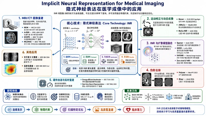
How can we recover reliable medical images from incomplete measurements affected by motion or artifacts? We combine implicit neural representations, imaging physics, and generative priors for MRI, CT, and X-ray reconstruction. Beyond visual quality, we focus on agreement with measured data and adaptability to changes in sampling, acquisition geometry, and hardware.
Developmental Imaging and Quantitative Analysis
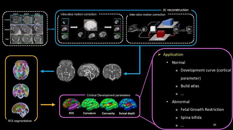
From fetal development to adult brain structure, we study how medical images can provide comparable, quantitative anatomical information. Our work centers on fetal MRI reconstruction, continuous developmental atlases, and quantitative brain analysis, with extensions to fetal body atlases, pathology, and clinical imaging. We connect image reconstruction with biomedical questions while preserving both population trends and individual variation.
Self-Supervised Denoising and Image Enhancement
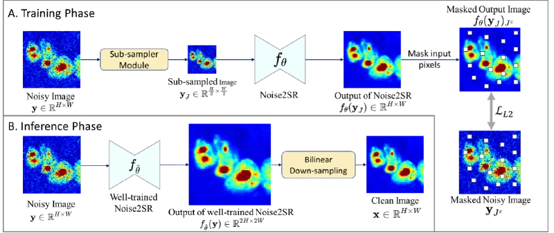
Low signal-to-noise ratio, limited resolution, and unknown intensity mappings affect both image detail and quantitative information. We study restoration without paired reference images through two complementary approaches: self-supervision from individual noisy images, and pretrained priors combined with degradation modeling. Our work spans MRI, fluorescence and electron microscopy, and blind inverse problems with unknown dynamic range compression.
Anatomical Modeling and 3D Physical Understanding
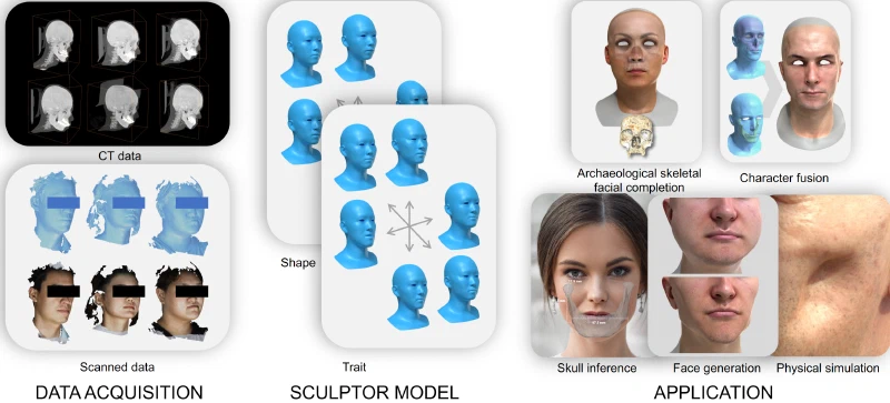
A 3D model should describe more than surface shape: it should also capture internal structures and their relationships. Using CT, MRI, and X-rays, we study joint bone–soft-tissue modeling, parametric hand representations, and facial shape prediction after orthognathic surgery. Extending this focus from appearance to internal structure, we also study density fields of real-world objects for physical property estimation and interaction.